What is XRayography?
The use of X-rays in art, is surprisingly enough, not new. In fact, it is part of the rich history of photography that started almost two hundred years ago.
Photography, as the name suggests, is essentially the act of drawing with photons. Its origins lie in the desire for two-dimensional artists to improve upon art and to directly imprint images with exposure to light. Before the discovery of x-rays, light and photons were considered different entities and the "wave vs. particle" theories had not yet been settled. The first photographic image was made in 1826 by a Frenchman named Joseph Niépce (who patriotically changed his name during the French Revolution to Nicéphore Niépce, the name that he is today known under as the inventor of the internal combustion engine), although the first usable image made with light (i.e. photons) was made by Daguerre in 1839. Daguerreotypes, as they are now known, were used for portraits, landscapes, documentation, and even scientific subjects. The ability to make colored photos did not exist then, so they were often painted in to simulate color. Since color photography is a rather recent discovery, this practice was carried on well into the 20th c. as well. Something that many of us will remember from our childhood is the painting of postcards with translucent paints so that the black and white photograph behind the paint would show through.

Louis Jacques Mande Daguerre (1787-1851)

Daguerrotype taken by Daguerre (1851)

Painted Postcard (1932)
In 1875 the process of capturing light onto a photographic medium had reached a level where the medium was sensitive enough to capture multiple images of a galloping horse giving rise to the possibility of simulating moving pictures. Unfortunately, however, it is science, in particular science for the purpose of gaining a military advantage over one’s enemy that drives new discoveries. Hence, photographic research was most often constrained to military, scientific and medical uses and the use of photography for artistic purposes did not much benefit from scientific support. As the battlefield saw many casualties, the need for a camera that could see inside the body – to set bones, find shrapnel, etc. – was needed.
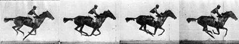
Sequential images of the same galloping horse showing the rapid succession of captured images
It just so happened that that two new discoveries were made in 1895 by a gentleman named Roentgen. He discovered that photographic emulsions are sensitive not only to regular light, but to light traveling at other frequencies, especially x-rays. He was interested in x-rays in particular because he had also discovered that these could traverse organic matter like human skin. Since the human body has different densities (skin, bones, cartilage), the differences in them could then be captured in gradations by the photographic medium behind the subject. These discoveries were of paramount importance to non-military scientific and cultural progress:
- Doctors could now determine the severity of an ulcer or cancer without having to operate or could locate a hairline fractures in a tibia.
- Scientists could now look deeper than with a microscope and could affect the chemical properties of matter.
- And finally, artists now had a whole new art form to explore. It opened the door to a whole new dimension in art, one who's surface has just been scratched with x-rays and that could witness far more development with other forms of light. For example, what if one could make an x-ray that could not only reveal the inside structure of an organism but also slightly change the chemical composition of the organism in the process and all this was captured by the photographic medium?
In 1905 Einstein showed that X-ray photons were actually similar in properties to light but modified to undulate at a much higher frequency. Simplified, the process could be described as directing light through an object to see its inner structure, essentially ‘drawing with light.' Viewed in this light (pardon the play on words), there is nothing strange or outrageous about x-rays.
Wilhelm Konrad Roentgen
(1845-1923)

Crookes Vacuum Tube typical of the type used by Roentgen

The world's first x-ray: Roentgen's wife, Bertha's hand

Albert Einstein
(Early 20th c. photo)
In 1925, scientists who had access to what were then astronomically priced government-owned x-ray machines, developed a few images for personal artistic purposes. True to the scientific mind, or perhaps due to pressing other concerns, these were not sold or marketed and were kept merely as ‘office art.’ Occasionally this is still done by doctors and scientists today. Then, in the early 1930’s, one such scientist, dr. Dain Tasker, a pioneer in the use of x-rays as art, made beautiful images of flowers. This was quite unique, but in the turbulent years that followed, the art was overshadowed by other events and so their use as art was forgotten for a while. Ironically, when these same images were found again, they were hailed as a historical milestone and auctioned off in New York for over $25,000 each.
X-ray by Dr. Dain Tasker
(1930)

'Positive' developed from
the x-ray at left by Tasker
Bearded Iris by Tasker
(1931)
There are a few other artists today who use x-ray machines to develop images of flowers. They are often dentists or doctors who have access to such equipment. Some are even satisfied in just using the x-ray negative (similar to those used in hospitals to set bones) as the art itself… Albert’s x-rayography is very different. He composes his images poetically by positioning the flowers or shells in ways that tell a story or remind one of an adage. He only uses the x-ray ‘negative’ to develop the ‘positive,’ using a specialized projector that he designed just for this purpose. Once developed, he only keeps the best pieces, the ones without flaws, somber patches, or over-exposures – sometimes only one in ten images is usable. Then he paints the composition in with the same translucent paints used over a century ago on daguerreotypes and postcards. The result is a beautiful union between passionate art and regimented science, monochrome depth and colorful surfaces, philosophical insight and pragmatic distance....
On a purely pragmatically level, the result is also an image that will not fade like a painting, or discolor over time like a computer-generated print. Each piece is a true painted photograph, one of only fifty of each type. After fifty are produced, the x-ray is retired and no new photographs are made from that negative. Like Roentgen and Tasker’s x-rays, they will provide centuries of beauty and value to their owner, and piece of the historical record of art history.
Sources:
Daguerre photos: http://www.rleggat.com/photohistory/history/daguerr.htm
Painted postcard: http://www.library.miami.edu
Galloping Horse: http://bowlingsite.mcf.com/Movement/HGal.html
Roentgen Images: http://www.softcode.com/X_ray.html
Eistein Photo: http://www.search.caltech.edu/Archives/action.lasso
Images by Dr. Dain Tasker: http://www.artland.com/cgi-local/SoftCart.exe/online-store/scstore/artist/?E+scstore
and http://www.artline.com/associations/ipa/show/show2001/exhibitors/greenberg/greenberg.html